
Chip-based modeling of mechanical skeletal muscle contractions

Chip-based modeling of mechanical skeletal muscle contractions

Delivery institution and unit

PhyMedExp INSERM U1046 – CNRS UMR 9214

Internship description

​

​

This innovative and interdisciplinary research project combines microsystems engineering, bioelectronics, and tissue culture to develop an organ-on-a-chip, or microphysiological system (MPS), modeling skeletal muscle contraction under physiological and pathophysiological conditions. Using human skeletal muscle biopsies taken from cohorts of healthy patients, the scientific challenge lies in recreating muscle microtissue in vitro to reproduce the complexity of this organ’s physiology. The longer-term objective is to use this same device in patients with acquired myopathy associated with organ dysfunction*, and thus compare the two states, from the molecular to the tissue level.

Study field of the internship

Disciplines

pre-requisite

Required study level

Other pre-requisite

Second-year Master’s internship – 6 months, from January to June 2027. The student must have a background in biology, specifically in cell biology (experience with cell culture and microscopy is an asset), as well as an interest in physics and electronics. The project requires the student to work across two different laboratories, adapting between a physics/electronics-oriented approach and its application to biological research.

Time period of the internship

6 months, January-June 2027

Application deadline

December 2026

Is the internship paid?

€4.50 per hour, which comes to €630–€650 per month

Is there accomodation support?

no

Other relevant informations

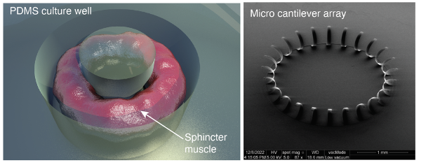
Our initial approach involves modeling an annular muscle (e.g., a sphincter) to test our ability to design molds in PDMS, and to adapt the integration of multiple and/or multiparameter sensor and/or actuator technologies for monitoring normal and then pathological biological processes. More specifically, we will develop culture wells in a silicone-based polymer (PDMS) using 3D-printed devices to create toroidal culture environments for generating sphincter-shaped muscle models. These structures allow muscle contraction around the central pillar and the measurement of these contractions by vacuum microscopy. We plan to record a homogeneous contraction phenomenon in healthy cases and a desynchronization of this same phenomenon in the pathological configuration.
In a second phase, we plan to integrate into these culture wells a microlever array like those developed by the IES team to measure the internal electrophysiology of organoids. These microlevers are also equipped with flexural sensors. Using a radial arrangement of the microlevers, placed in a toroidal culture well, we plan to investigate the possibility of stimulating and recording the electrical activity of sphincters as well as their mechanical contractions.
The student recruited for this work will participate in cell culture techniques (isolation, differentiation, and manipulation of fibroblasts and myoblasts) to prepare tissue-on-a-chip models. She/he will also participate, concurrently, in the IES laboratory, in the development of PDMS molds to model organoids according to predefined models (sphincter/microlever). Finally, this six-month period will include initial electrical stimulation trials using microfluidic technologies and microelectrode arrays (MEAs) to record the first responses of these advanced cell models.
Our long-term perspective is based on a multi-organ approach to understanding and treating systemic or inter-organ diseases, notably through the integration of other tissues (e.g., nervous)

Interested?
In order to apply please contact the internship's responsible :

Eric ESTEVE